
What Exactly is ALK Positive Lung Cancer?
Find out more about the history of ALK fusion with an emphasis on early discovery, diagnostic methods, and the development of the first targeted ALK inhibitors.
ALK Lung Cancer 101
These links provide a good general explanation of what ALK-positive lung cancer means for a newly-diagnosed patient.
https://lcfamerica.org/lung-cancer-info/types-lung-cancer/alk-positive-lung-cancer/
https://www.webmd.com/lung-cancer/faq-stage-four-lung-cancer-with-alk#1
How did it all start?
In 1996, a group of scientists in Japan wanted to find out a common factor for lung cancer patients that would allow them to treat this disease more effectively. They used an adenocarcinoma biopsy sample from a 62-year old, smoking, lung cancer patient for their research. They isolated one alteration where there is a fusion of two genes (EML4 and ALK genes) from chromosome 2. The fusion product has also been found before in lymphomas and inflammatory myofibroblastic tumors. This group demonstrated that EML4-ALK fusion alone can induce a tumor formation in mouse models. The authors then went back into the 33 patient samples they had available and found three more patients showing this EML4-ALK variant 1 configuration. They also found other variations of ELM-4 ALK fusion, named variant 2. The ELM-4 ALK fusion change does not seem to overlap with the known EGFR mutations that also cause lung cancer. They speculated that researchers can target the cancer by reducing the activity of this ELM-4 ALK fusion protein.
https://www.nature.com/articles/nature05945
In 2010, a review was done in Europe. By then, many research groups had worked on the biology and treatment of ELM4-ALK lung cancer. There is a distinct look to these cancer cells, looking more like gastric, colonic, and breast adenocarcinomas. There are numerous variations of the EML4-ALK fusion. In addition, it has been reported that various different genes can be fused with the ALK gene.
The main detection methods for a patient to be diagnosed with the ELM4-ALK are: 1) FISH (fluorescence in-situ hybridization); 2) and IHC (immunohistochemical) analysis.
FISH can be quickly summarized as follows: A gene is labelled 5’ and 3’, where 5’ is the “head” of the gene and the 3’ is the "tail" of the gene. A green and an orange tag with two probles are then placed for the ALK gene, one for the 5’ head and one for another segment in the middle of the ALK gene. A biopsy sample is treated with the color-tagged probes and the technicians can visually see the two dots. If the ALK gene is connected in its proper configuration, the green and orange dots are lined up properly. If the ALK gene has changed in any way, such as through inversions, fusions, or even deletions, then the two dots are not in regular configuration.
For IHC, the biopsy sample is sliced thinly and stained with antibodies specific to the ALK protein. However, this method only detected 4 out of 10 ALK rearrangements/fusions detected by the FISH method.
The ALK protein naturally functions as a kinase. This means that it takes away a phosphate group from a protein. This opens up a logical treatment venue to generate an inhibitor to the ALK protein. The molecule called PF-02341066 (Crizotinib or its brand name, Xalkori) was the first one to show very promising results.
https://www.ncbi.nlm.nih.gov/pmc/articles/PMC2888755/
By 2014, results were available from an open-label, phase 3 trial comparing ALK inhibitor Crizotinib against the standard chemotherapy for advanced ALK-positive non-small cell-lung cancer (NSCLC). The treatments consisted of either 250mg of Crizotinib twice per day or Pemetrexed plus Cisplatin or Carboplatin every 3 weeks for 6 cycles. If there was disease progression under chemotherapy, the patients were allowed to switch to the Crizotinib protocol. The result showed a significant increase in progression free survival of 10.9 months with Crizotinib vs. 7.0 months with standard chemotherapy. In addition, most patients reported better quality of life with Crizotinib.
https://www.nejm.org/doi/full/10.1056/nejmoa1408440
A simplified biological diagram of ALK function in the cell
ALK wild-type means the normal, unaltered ALK gene in our bodies. This functions normally as a kinase and is anchored onto the cell membrane. Normally, there would be a protein floating outside of the cell wall to trigger or activate the ALK protein to take a phosphate from its substrate. However, when ALK fuses with another molecule such as EML4, ALK loses its anchor to the cell membrane and the trigger to turn the kinase function on or off. The “floating ALK” pictured below is aberrant in its function, which means unable to be turned off properly. This activated ALK-fusion protein turns on lots of downstream signal pathways which allow the cell to continue to grow uncontrolled.
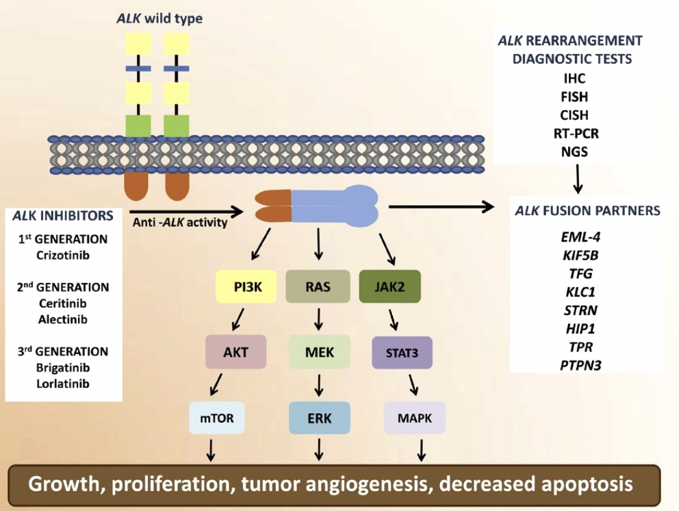
More methods for diagnosing ALK-fusion/rearrangement have been developed in recent years. IHC and FISH have been described above in more detail. CISH is "chromogenic in-situ hybridization". This method is very similar to FISH, however the tag is color-based and is cheaper to detect when compared to the fluorescence tag involved in FISH. RT-PCR is "reverse-transcriptase polymorphic chain reaction" where a sample of RNA from the cells is transcribed into DNA and amplified to detect a segment. In this case, a specific fusion product would be identified. NGS, "next generation sequencing", is a machine-driven, simultaneous read of a segment. Color is used as a technique to sequence small strands of RNA or DNA.
We apologize that the proper reference for the image has been accidentally deleted.
Go beyond the basics in our next blog: ALK and the MET Amplification and TP 53 HERE